"One problem chicken is enough to take out an entire flock of birds," Chet said at the beginning of class. Without proper maintenance (cleaning, feeding, etc) and up-to-date vaccinations that could mean a loss of revenue and in turn, the loss of a job. At the UIUC poultry farms, all birds in Chet's flock are up-to-date on vaccines and are given feed and water easily through the use of mechanized system using feed and water lines that are installed through the cage system.
Now, because all of the birds at these farms have already been vaccinated, Chet taught us how to properly vaccinate a chicken by demonstrating the same methods he would use to vaccinate a chicken, but through the use of a blue-dye (the mixing agent for real vaccine that has no actual effect) instead of a live vaccine. First,
he laid out a table with a handbook of all vaccinations required by the government, a stabber (a small tool with two, long, and of medium size metal rods with metal pointy ends, both of which had wells in the middle of them) and a pricker (a sharp small rod for puncturing chicken veins to release blood with a loop at the opposite end to collect it). Next, he took the stabber, dipped it into the loading dye and tapped it against the edge of the bottle to remove excess dye. Chet paused here to explain the importance behind tapping the stabber: Each vaccine is expense and comes in limited supply, so using this technique will maximize the amount of birds you can vaccine. Then, he took a chicken, spread one of its wings out and punctured its wing web (the same place where chickens are wing-banded) with the stabber and then immediately removed it. "There's one vaccinated chicken," he said to the class as he spread the wing out to show a medium-size blue circle that appeared around the stab-wound.
Students were then instructed to retrieve a chicken from the cages and return to the area to begin fake administration of vaccines as well as perform Pullorum testings. After each student successfully did each of these things, we all moved to the specialized laboratory to dissect different aged chicks and chickens.
The first thing we all noticed when we walked in and circled-up was three very sickly looking chickens sitting on the floor. Pam Utterback, Chet's wife, led the discussion of this dissection section as well as the introduction to this section which regarding proper techniques to euthanize chickens. Pam explained that there were 3 types of euthanasia that the farms practiced: Carbon Dioxide gassing, electrocution, and cervical dislocation. The first two are commonly used to cull groups of chickens, so the technique that the class was going to be taught how to use was cervical dislocation. Pam stated that she was certified to teach anyone how to do perform this procedure so if anyone wanted to volunteer and practice on a dead-bird they were more than welcome to. Chet jumped in and stated that if anyone was timid that they should think of it like this, "Would you like to have the air removed from the space around you, have electricity run through your body at an extremely high voltage, OR have a ninja come and karate-chop you in the neck so that you die instantly?" Out of the 3-classes I assisted in, only 1 of the classes had students brave enough to attempt this procedure after Chet's encouraging speech.
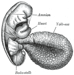
Following the euthanasia of these 3 cull of the flock, all the students circled around a large table and then Pam placed 1-day old and 3-day old chicks in front of each student. These chicks were chosen to demonstrate how chicks are able to live through being shipped across countries (about 3 days) without being fed any food or water. This is due to the fact that chicks retain something called the 'yolk sac' inside them as a nutrient source. On the day 1 chicks the sac is very large, however, on the day 3 chick the sac has almost entirely disappeared. Following these chicks, the each student in the class was given an adult culled chicken. We dissected each chicken to see both the general outline/picture of a chickens digestive system, as well as
potential reasons behind why they died.
A majority of the chickens that were culled had been used for ovarian cancer research and their cause of death was because of ovarian cancer. Inside of them, we were shown their ovaries and almost all of them had developed uterine or ovarian cysts on their ovaries. Compared to normal ovaries, cancerous ovaries look cauliflower like and cause the chickens problems with producing eggs as these ovaries are now more/fully obstructed and disease ridden.Following dissection of hens, Pam dissected a rooster and showed us the difference in physique, disease, and sex structure of the male and female chicken. The only big differences noted were that the roosters tended to have more meat (which makes sense as they will grow bigger then hens) and the size of the testicles compared to the size of the ovaries.
After dissection, Chet showed us his collection of weird things he has collected over the years from these multiple dissections. A lot of the things he collected were unusual ovarian cysts and odd, un-coated eggs, but the one that stood out most to me was something that could be collected after every dissection and that was of the brain compared to the chickens eyes. I was amazed to see that the brain was smaller than one of the chicken's eyes, absolutely incredible.


No comments:
Post a Comment